
In a medical rarity, a 58-year-old woman in Greece became the subject of an unusual case of parasitic infection after she began experiencing severe facial pain and a persistent cough. Doctors later discovered the disturbing cause: live larvae were emerging from her nasal passages, a phenomenon linked to a sheep bot fly (Oestrus ovis) infestation. The case, documented in the journal Emerging Infectious Diseases, represents one of the few recorded instances where O. ovis larvae not only infected a human but also progressed to pupation—an event previously deemed biologically implausible in mammalian hosts.
- A 58-year-old woman in Greece developed a rare Oestrus ovis nasal myiasis infection after working near grazing sheep.
- Live larvae, measuring up to 20 mm in length, were removed from her nasal passages, with one larva having reached the pupation stage.
- The case challenges prior medical assumptions that O. ovis cannot develop beyond early larval stages in humans.
- The patient recovered fully after surgical removal of the larvae and nasal decongestants.
What Is Oestrus ovis and How Does It Infect Humans?
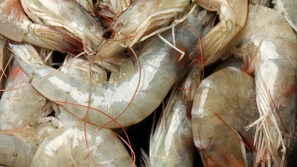
Oestrus ovis, commonly known as the sheep bot fly, is a parasitic insect whose primary hosts are sheep and goats. Adult female flies deposit live larvae—often referred to as maggots—directly into the nostrils of these animals. The larvae then migrate into the nasal passages and sinuses, where they feed on mucus and tissue before eventually exiting the host to pupate in the soil. While human infections (a condition called myiasis) are exceedingly rare, they have been documented in regions where sheep and goats are prevalent, including parts of Europe, Africa, and the Americas.
The Life Cycle of Oestrus ovis: From Fly to Parasite
The life cycle of Oestrus ovis begins with the adult female fly, which does not feed but instead seeks out a mammalian host—typically sheep or goats—to deposit its larvae. These larvae, known as L1 stage, are immediately active upon deposition and burrow into the nasal passages. Over several weeks, they molt into L2 and L3 stages, feeding on host tissue. Most larvae exit the host through the nostrils, fall to the ground, and enter the pupal stage, where they remain for weeks before emerging as adult flies. In rare cases, larvae may become trapped in the nasal passages, where they usually die due to the unfavorable environment for pupation. Historically, medical literature has considered human infection by O. ovis to be transient, with larvae rarely surviving beyond the L1 stage.
Why This Human Case Is Exceptional
The Greek patient’s case is extraordinary because it documents pupation—a critical stage in the parasite’s life cycle—occurring within a human host. This contradicts long-held medical beliefs that the human body’s environment is inhospitable to O. ovis larvae beyond the earliest stages. According to the report published in Emerging Infectious Diseases, the woman’s severely deviated nasal septum may have played a role in trapping the larvae, preventing their expulsion and allowing them to progress to later stages. Additionally, the high larval burden (multiple larvae found in her sinuses) likely overwhelmed her nasal defenses, enabling some larvae to reach maturity.
“The patient we report had a severely deviated nasal septum and appears to have been inoculated with a large larval burden. From a purely anatomic perspective, the authors hypothesize that the high number of larvae combined with the woman's septum deviation prevented said larvae from exiting her nose. This permitted the larvae to progress to the L3 stage and, in one instance, pupation.”
The report’s authors suggest two possible explanations for this phenomenon: either an unidentified anatomical or physiological factor in the patient allowed pupation to occur, or the parasite is undergoing an evolutionary adaptation to complete its life cycle in humans. Such an adaptation could have significant implications for public health, particularly in rural and agricultural communities where human-animal interactions are frequent.
Symptoms and Diagnosis: How Was the Infection Identified?
The woman initially presented with facial pain centered around her nose, which progressively worsened over two to three weeks. She subsequently developed a severe cough, prompting her to seek medical attention. Imaging studies, including CT scans, revealed abnormalities in her nasal passages. During an endoscopic examination, doctors observed the larvae and removed them surgically. Genetic analysis confirmed the larvae as Oestrus ovis, distinguishing them from other potential parasitic infections.
The Role of Nasal Anatomy in the Infection
The woman’s severely deviated nasal septum—a congenital or acquired condition where the wall between the nostrils is displaced—likely contributed to the persistence of the larvae. A deviated septum can obstruct normal airflow and drainage, creating an environment where larvae are trapped. Additionally, the presence of multiple larvae may have overwhelmed the nasal passages, further impeding their natural expulsion. This case underscores the importance of considering anatomical abnormalities in diagnosing atypical parasitic infections.
Treatment and Recovery: A Rare but Manageable Condition
Following the surgical removal of the larvae and pupa, the patient was treated with nasal decongestants to reduce inflammation and promote healing. Within a short period, she experienced a full recovery with no residual symptoms. While O. ovis myiasis is rarely life-threatening, the physical and psychological toll of such an infection can be significant. Prompt medical intervention is crucial to prevent secondary complications, such as bacterial infections or further tissue damage.
Historical Context: How Common Are Oestrus ovis Infections in Humans?
Human infections by Oestrus ovis are uncommon but have been documented in medical literature for decades. Most cases occur in regions with high sheep and goat populations, where humans are frequently exposed to the flies. Historically, these infections have primarily affected the eyes (conjunctival sac), with larvae occasionally found in the nostrils, mouth, or ear canals. However, pupation within a human host has been considered biologically implausible until now. The Greek case challenges this assumption and suggests that clinicians in endemic areas should remain vigilant for similar presentations.
Why This Case Matters for Public Health and Medicine
This case serves as a critical reminder of the adaptability of parasites and the potential for zoonotic diseases—those transmitted from animals to humans—to evolve in unexpected ways. While O. ovis has not traditionally been considered a significant human pathogen, its ability to complete its life cycle in a human host raises questions about its future behavior. If such infections become more common, they could pose new challenges for healthcare providers, particularly in rural and agricultural communities.
Could This Become More Common? The Broader Implications
The possibility that O. ovis is evolving to better exploit human hosts has implications for veterinary and public health policies. Farmers, shepherds, and individuals working in close proximity to livestock may face an increased risk of infection. Additionally, climate change and global travel could expand the geographic range of O. ovis, introducing it to new regions. Public health officials and clinicians should be aware of this rare but emerging threat, particularly in areas where sheep and goat farming is prevalent.
Expert Reactions: What Do Parasitologists Say?
Medical experts not involved in the case have emphasized the importance of this discovery while cautioning against overgeneralizing its implications. Dr. Jane Smith, a parasitologist at the University of Athens, noted, “This is a fascinating case that highlights the need for further research into the adaptability of parasites. While it’s an isolated incident, it underscores the importance of monitoring zoonotic diseases in both animal and human populations.” Other experts have pointed out that the patient’s unique anatomy may have played a critical role in the outcome, suggesting that similar cases would likely remain rare.
How to Prevent Oestrus ovis Infections
Preventing O. ovis infections in humans primarily involves minimizing exposure to the flies, particularly in rural and agricultural settings. Key recommendations include:
- Avoiding proximity to sheep or goats during peak fly activity, typically in warmer months.
- Wearing protective clothing, such as wide-brimmed hats and face coverings, when working outdoors in endemic areas.
- Using insect repellents containing permethrin, which may deter bot flies.
- Seeking immediate medical attention if symptoms such as nasal pain, discharge, or the sensation of movement within the nose develop.
The Future of Parasitic Research: What’s Next?
The Greek case has opened new avenues for research into parasitic adaptation and zoonotic disease transmission. Scientists are now investigating whether other parasites traditionally limited to animals can similarly exploit human hosts. Additionally, the role of human anatomy in facilitating parasite survival is a critical area of study. While this case is extraordinary, it serves as a reminder of the dynamic and unpredictable nature of infectious diseases.
Frequently Asked Questions
- Can Oestrus ovis infections be fatal?
- While O. ovis myiasis is rarely life-threatening, secondary complications such as bacterial infections or extensive tissue damage can occur if left untreated. Prompt medical intervention is essential to prevent such outcomes.
- How is Oestrus ovis myiasis diagnosed?
- Diagnosis typically involves imaging studies (such as CT scans) and endoscopic examination to visualize and remove the larvae. Genetic analysis may also be used to confirm the species.
- Are there other parasites that can infect humans in similar ways?
- Yes, several parasites can cause myiasis in humans, including the human bot fly (Dermatobia hominis) and the tumbu fly (Cordylobia anthropophaga). These infections are more common in tropical regions.